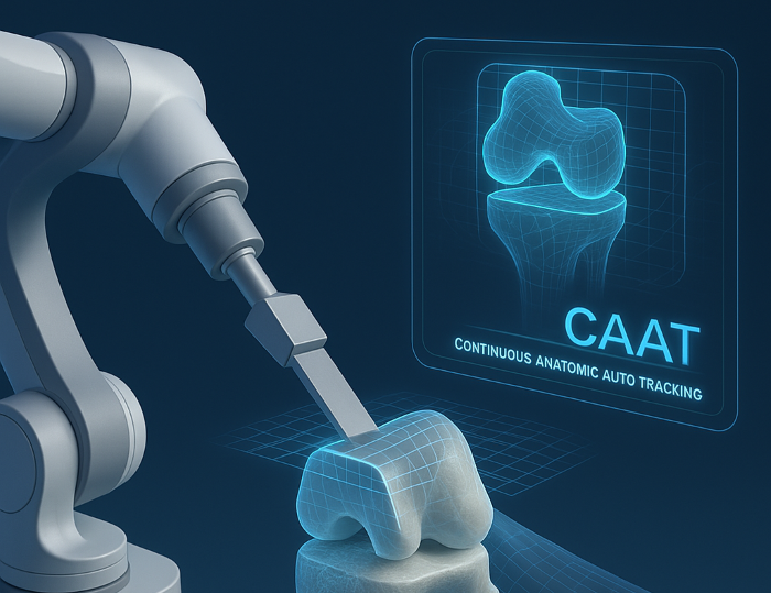
You’ve relied on hardware-based navigation methods and multi-step registration workflows for decades. Now there’s a real alternative: complete knee resections executed without any invasive hardware. Using spatial computing for continuous anatomical tracking, surgeons can guide robotic saws purely by vision, eliminating bone-pinned trackers entirely.
This method was demonstrated in cadaveric resections using the CAAT platform, Continuous Anatomic Auto Tracking, developed by Visie. The system delivers tracking resolution comparable to infrared-based navigation yet requires no rigid fixation to bone. The resections were completed with only a scanner and robot arm operating in sync. This development changes what it means to “track anatomy” during surgery and raises real questions about whether physical reference arrays still belong in the operating room.
How visual tracking replaces hardware
Traditional computer‐assisted systems begin with registration of landmarks via pins or palpable markers, aligning a digital map to patient anatomy. Then trackers monitor motion during surgery. Now marker‐less optical tracking registers the distal femur and proximal tibia in a split second and tracks them continuously in real time as they move during cutting. Spatial computing interprets visible anatomy and maintains alignment between anatomy and robot. Cuts involving the anterior surface, posterior cortex, distal femoral plane, and posterior chamfer are all executed using only computed spatial data. Manual registration is not required.
Why no trackers matters to you
Tracker pins raise risk of fractures, added trauma, infection sites, and extra operating time. Removing arrays simplifies the setup and reduces the need for rigid fixation points. Without invasive tracking, you maintain sterile field flexibility. Precision remains intact because it is delivered optically instead of mechanically.
Trends in navigation: Toward optical, infrastructure free systems
Recent research supports these approaches beyond TKA. Visual markerless tracking using RGB D cameras can achieve approximately 2 to 4 mm bone accuracy even with occlusions, and it avoids additional hardware entirely. For ACL reconstruction, dynamic arthroscopic systems running at 25 fps with sub 40 ms latency use multi-level memory models to track anatomy without external fiducials. Their localization error drops by approximately 45 percent compared with static methods. Ultrasound based tracking powered by deep learning demonstrates sub millimeter precision from raw signal data alone. This occurs without motion capture or markers and does not require any third-party external tools. All signs point to surgical navigation moving toward zero infrastructure computing.
What’s ahead for orthopaedic surgery
You’re at a point where tracking arrays may become optional rather than mandatory. It’s plausible that in the near future robotic systems for TKA will rely on vision platforms for bone registration and cutting alone. That approach may expand to spine, cranial, upper limb, and foot and ankle procedures whenever anatomy is exposed. Real time cartilage and bone stock mapping may add dynamic input to soft tissue balancing and deeper alignment decisions dynamically as cutting proceeds.
Integrating optical tracking with existing robotic platforms could make navigation more universal. Instead of rigid device specific arrays, you’d pair any compatible scanner with a compatible robot. Surgical planning and intra operative decision making could be more fluid when anatomy is tracked optically, especially in imageless or CT based workflows.
Surgeons’ perspective: Impact on workflow and accuracy
You can streamline the procedure by reducing preparation steps and skipping array placement. You still preserve or exceed current accuracy benchmarks. Vision based guidance may open up lower-profile incisions, reduce tissue manipulation, or eliminate the need for fixed reference points in specific workflows. Technologies like CAAT serve as proof that optical tracking can meet clinical standards for precision without invasive hardware.
Navigating toward a cleaner, faster future
Surgeons are already using spatial computing to guide bone cuts in total knee arthroplasty without depending on tracking pins or rigid fixation points. The system operates without physical infrastructure and functions independently of any hardware attached to the patient. That reality forces a question: If accurate resections can be performed under those conditions, what role should traditional tracking systems still serve?
The shift away from physical reference tools is already underway in practice. Optical navigation is beginning to change how surgeons approach alignment and control during procedures. For those using conventional methods, this may be the right time to evaluate whether those tools still offer the most efficient path to precision.
Disclaimer: This article is intended for informational purposes only and does not constitute medical advice. The technologies and techniques discussed herein, including markerless optical tracking and the CAAT platform, may not be approved or available in all clinical settings. Surgeons and healthcare providers should consult relevant clinical guidelines, peer-reviewed research, and device manufacturers before adopting new surgical tools or altering treatment protocols. Individual patient needs and professional judgment should guide all clinical decisions.
Sources
Deep Learning based acoustic measurement approach for robotic applications on orthopedics
Occlusion-robust Visual Markerless Bone Tracking for Computer-Assisted Orthopaedic Surgery
VISIE Announces Continuous Anatomic Auto Tracking (CAAT) for RA-TKA
VISIE Executes First Pin-Free Knee Bone Cuts Using Spatial Computing


