Key takeaways:
- Evidence remains mostly preclinical. Printing enables patient-specific constructs that reproduce cartilage over bone by layering materials under CAD control.
- Hybrid designs that pair bioactive hydrogels with supportive lattices or cartilage-zone ECM have improved mechanics and early in vivo readouts in animal models.
- Clinical use remains distant, but preclinical designs are identifying features that may guide future surgical applications.
Why the osteochondral unit keeps failing traditional fixes
The osteochondral unit poses unique challenges. Cartilage is aneural and avascular. It must integrate with vascularized subchondral bone across a calcified interface. This mismatch undermines durability after marrow stimulation or mosaicplasty. The interface is consistently identified as the failure point. Despite remaining largely preclinical, 3D-bioprinted osteochondral grafts offer a novel design strategy that mimics native tissue by continuously integrating mechanical behavior with spatially organized biological activity.
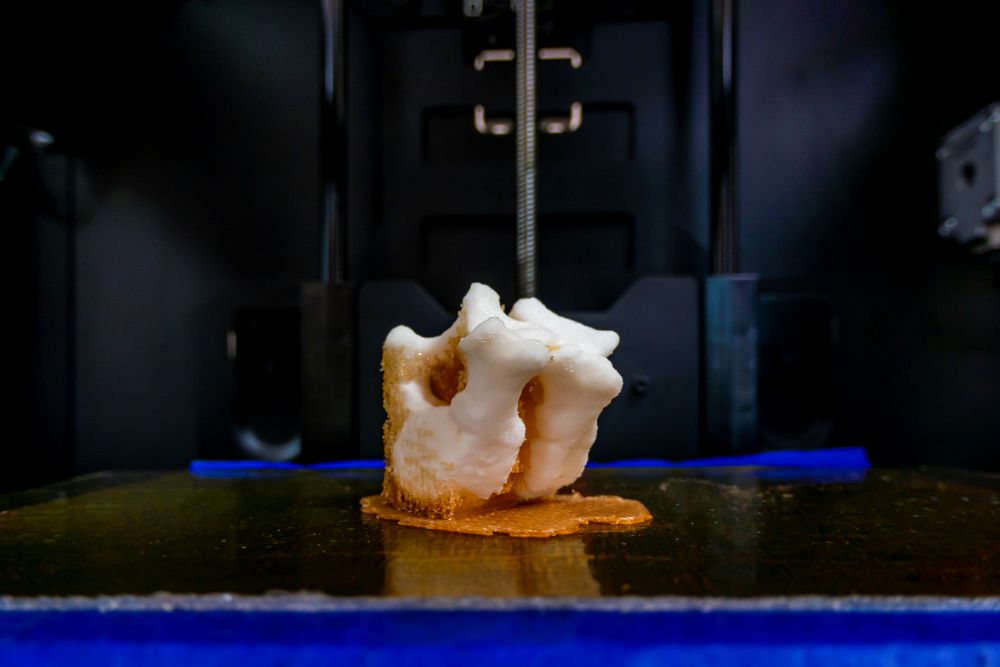
What bioprinting actually adds
Bioprinting enables zonal grafts that behave more like native tissue than uniform plugs. By varying stiffness, composition, cell content, and architecture through depth, a single construct can withstand compressive loads at the surface while promoting bone integration beneath. CAD-guided design also makes defect-specific grafts possible, matching geometry and engineered porosity to the surgical site with a precision stock implants cannot achieve. The next challenge is translating this design control into reproducible, clinically reliable constructs.
Scaffolds and bioinks in current designs
Bioprinting approaches generally fall into two categories: Hydrogels for the cartilage zone and reinforced composites for the bone zone, each designed to meet different biological and mechanical demands.
Hydrogels dominate the chondral phase because they can be printed at low temperatures that preserve cell viability while supporting high densities of chondrocytes or progenitor cells. Hyaluronic acid and gelatin derivatives are among the most frequently used carriers, offering a biologically familiar environment for cartilage formation. Researchers are now using decellularized cartilage ECM as a printable bioink to provide native biochemical cues and promote cartilage-like repair.
For the osseous phase, composite designs that combine natural polymers with synthetic frameworks are increasingly favored. These hybrids balance bioactivity with the structural reinforcement needed to withstand load, making them more suitable for the mechanical requirements of subchondral bone.
Mechanical limitations and the role of hybrid designs
For load-bearing osteochondral defects, inadequate mechanical strength is the primary reason hydrogel-only constructs fail before integration. Reinforcement is therefore essential.
A 2024 study illustrated this with a biphasic plug: A hyaluronic-acid hydrogel formed the cartilage layer, while a GelMA-based bone zone was co-printed with a biodegradable thermoplastic-ceramic lattice. This reinforcement improved construct strength during culture while preserving cell viability and lineage-specific differentiation. Although the reported modulus remained below that of native cartilage, it marked a significant improvement over hydrogel alone.
These findings highlight a consistent theme across recent work: Hybrid scaffolds that combine bioactive hydrogels with supportive lattices are the most promising route toward clinically viable osteochondral grafts.
Advances in cartilage-zone signaling
A recent in vivo rabbit study showed that embedding decellularized cartilage ECM in the chondral zone led to more complete defect fill and higher histological cartilage scores at six weeks. In preclinical models, gradient scaffolds that transition from cartilage to bone composition consistently outperformed uniform designs. Still, confirmation in larger animal and clinical studies is needed before these strategies can be considered durable solutions.
Vesicle-based signaling beyond growth factors
Extracellular vesicles derived from mesenchymal cells are now recognized as bioactive cargo with potential advantages over traditional growth factors. In rodent bone-defect models, vesicle-loaded hydrogels enhanced osteogenesis and angiogenesis, both of which are critical for maintaining subchondral support beneath a chondral cap. Recent studies emphasize controlled delivery of zone-specific signals within printed scaffolds and position vesicles as a promising alternative to conventional growth factors.
Fit and intraoperative reality
Patient-specific grafts are technically feasible because CAD-based design can match the shape and internal gradients of a defect with far greater fidelity than stock implants. For surgeons, that precision could improve fit and integration, but it also raises practical questions: Will the grafts arrive sterile and ready to implant? Will their mechanical properties be consistent from batch to batch? Can manufacturers define standardized benchmarks for multi-material constructs that regulators and surgeons can trust? Until reproducibility and regulatory pathways are resolved, defect-specific fit remains promising but not yet practical for clinical use.
Where the evidence stands today
Clinical trials are not yet available. Most data still come from animal models and bench testing that measure mechanical performance and tissue integration. Even so, the field has progressed beyond proof-of-concept. Current designs include hybrid plugs that preserve strength under early load and cartilage-zone ECM that recruits host cells while encouraging matrix production. CAD-shaped grafts tailored to individual defects are also in development. These approaches mark a step forward compared with earlier uniform constructs.
For surgeons, the outlook is gradual but positive. Bioprinting is still on the bench, but it is solving the exact problems that make osteochondral repair unreliable today. If these designs hold up in large-animal and eventual human trials, surgeons may finally gain access to grafts that do more than buy time. They could deliver lasting restoration of the cartilage-bone unit.
Sources
3D bioprinted scaffolds for osteochondral regeneration: advancements and applications
3D bioprinting techniques and hydrogels for osteochondral integration regeneration
3D printed osteochondral scaffolds: design strategies, present applications and future perspectives
Characterization of biological and mechanical properties of 3D-bioprinted osteochondral plugs
Decellularized cartilage tissue bioink formulation for osteochondral graft development
Progress in 3D Bioprinting Technology for Osteochondral Regeneration


